


化工儀器網(wǎng)>產(chǎn)品展廳>醫(yī)療器械設(shè)備>血液學(xué)分析設(shè)備>流式細胞儀>Cell volume measurement Cell volume assay實時跟蹤貼壁細胞體積


Cell volume measurement Cell volume assay實時跟蹤貼壁細胞體積
- 公司名稱 世聯(lián)博研(北京)科技有限公司
- 品牌 其他品牌
- 型號 Cell volume measurement
- 產(chǎn)地
- 廠商性質(zhì) 代理商
- 更新時間 2020/2/27 23:14:19
- 訪問次數(shù) 624
Cell volume assay實時跟蹤貼壁細胞體積細胞力學(xué)細胞體積Cell volume measurem
聯(lián)系方式:李勝亮13261877206 查看聯(lián)系方式
聯(lián)系我們時請說明是化工儀器網(wǎng)上看到的信息,,謝謝!
細胞應(yīng)力加載儀,3細胞打印機,NanoTweezer新型激光光鑷系統(tǒng),PicoTwist磁鑷,美國NeuroIndx品牌Kuiqpick單細胞捕獲切割系統(tǒng)
| 產(chǎn)地類別 | 進口 | 產(chǎn)品種類 | 細胞分析系統(tǒng) |
|---|---|---|---|
| 價格區(qū)間 | 面議 | 應(yīng)用領(lǐng)域 | 醫(yī)療衛(wèi)生,生物產(chǎn)業(yè) |
Cell volume assay實時跟蹤貼壁細胞體積
Cell volume assay
Tracking the cell volume of adherent cells in real time
4DCELL DEVICE
Cell volume measurement technology
READ-OUTS
Cell volume, ion pumps
STANDARD CULTURE LIMITATION
There are several physiological and pathological processes where cells undergo a change of volume. However, there are no reliable methods that can be applied to accurately measure volume of adherent cells in real time.
CELL VOLUME ASSAY BENEFITS
Cells are cultured in an optically transparent chamber that enables to accurately determine their volume along time and to follow in parallel the biochemical processes responsible for the volume change, as for example activation of ion pumps.
EXAMPLES
Volume tracking from interphase stage, to mitosis of Raji cells [2].
![]()
(A) Cells are placed in poly(dimethylsiloxan) chambers of calibrated height set by pillars, in medium supplemented with FITC-Dextran. Bottom picture: cells exclude ?uorescence on epi?uorescence images (Scale bar 20 mm).
(B) The ?uorescence pro?le corresponding to the dotted line in (A): maximum and minimum of ?uorescence intensity correspond to chamber maximal height (background) and zero height (pillar), respectively. Right: these values are used to calibrate the signal and calculate the optical thickness of the cells.
(C) Finally, cell volume is obtained by integrating the total ?uorescence intensity over the cell area.
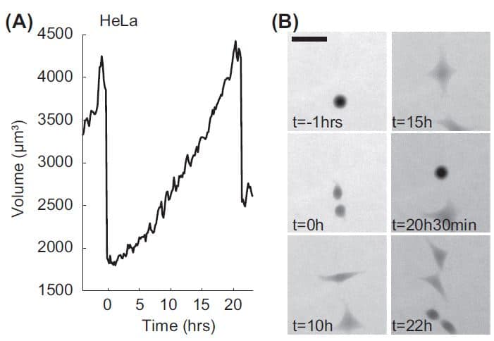
(A) Volume trajectory of a HeLa cell. The two volume overshoots at the beginning and the end correspond to transient volume increase in mitosis with the ?rst one corresponding to the mother cell and the second one to the daughter cell.
(B) Raw Fluorescence images of the cell in (A) with FXm. Scale bar 50 mm.
REFERENCES
[1] Zlotek-Zlotkiewicz, E. et al. (2015). Journal of Cell Biology, 211(4), 765–774.
[2] Cadart, C et al. (2017). Methods in Cell Biology, 139,103-120.


 采購中心
采購中心
 化工儀器網(wǎng)